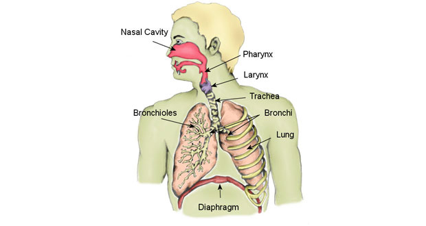
Organs of the Respiratory System
- The respiratory system is crucial for the exchange of gases between the body and the environment.
- It consists of various organs and structures that work together to facilitate breathing and gas exchange.
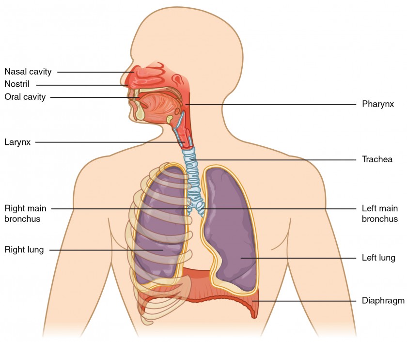
- Key components include the nose, pharynx, larynx, trachea, bronchi, lungs, and diaphragm.
1. Nose and Nasal Cavity
Functions:
- Acts as the primary entrance for air into the respiratory system.
- Filters out dust, pollen, and other particles from the air.
- Warms and humidifies the air to body temperature and moisture levels.
- Contains olfactory receptors for the sense of smell.
Anatomy:
- External nose: Composed of bone and cartilage, covered by skin.
- Nasal cavity: Hollow space behind the nose lined with mucous membranes and cilia.
- Nasal conchae: Bony projections that increase the surface area of the nasal cavity, aiding in air filtration and humidification.
- Nasal septum: Divides the nasal cavity into left and right sides.
2. Pharynx (Throat)
Location: Located behind the nasal cavity and mouth, extending to the larynx and esophagus.
Functions:
- Serves as a common pathway for both air and food.
- Participates in swallowing reflexes.
- Contains tonsils, which are part of the body's immune system.
Sections of the Pharynx:
1. Nasopharynx: Located behind the nasal cavity, serves only as an air passageway.
2. Oropharynx: Located behind the mouth, serves as a passage for air, food, and drink.
3. Laryngopharynx: Lowest part of the pharynx, connects to both the esophagus and larynx.
3. Larynx (Voice Box)
Location: Located between the pharynx and trachea, at the level of the Adam's apple (thyroid cartilage).
Functions:
- Provides a passageway for air between the pharynx and trachea.
- Contains the vocal cords, which vibrate to produce sound during speech and singing.
- Acts as a protective mechanism during swallowing, closing off the airway to prevent food and drink from entering the lungs.
Cartilages of the Larynx:
- Thyroid cartilage: Forms the Adam's apple, protects the vocal cords.
- Epiglottis: Leaf-shaped cartilage that covers the opening of the larynx during swallowing, directing food and drink into the esophagus.
4. Trachea (Windpipe)
Location: Extends from the larynx to the bronchi, located anterior to the esophagus.
Structure:
- Composed of C-shaped rings of hyaline cartilage, which provide support and prevent collapse of the trachea during inhalation.
- Lined with mucous membranes and cilia, which trap and remove particles from the air.
- Divides into the left and right primary bronchi at the level of the fifth thoracic vertebra.
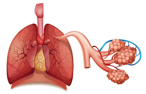
5. Bronchi and Bronchioles
Bronchi:
- Primary bronchi: Branch off from the trachea into the left and right lungs.
- Secondary bronchi: Enter each lobe of the lungs.
- Tertiary bronchi: Further divide within each lobe into smaller bronchioles.
Bronchioles:
- Smaller airways that lack cartilage support.
- Branch extensively within the lungs and eventually lead to the alveoli.
- Smooth muscle in bronchioles regulates airway diameter, influencing airflow.
6. Lungs
Location: Paired organs located in the thoracic cavity, on either side of the heart.
Structure:
- Each lung is enclosed by a double-layered pleural membrane, consisting of the visceral and parietal pleura.
- Divided into lobes: Right lung has three lobes (superior, middle, inferior), while the left lung has two lobes (superior, inferior) to accommodate the heart.
- Bronchial tree branches within the lungs, culminating in terminal bronchioles and alveolar ducts.
7. Alveoli
Structure:
- Tiny air sacs clustered at the end of bronchioles.
- Surrounded by a network of pulmonary capillaries.
- Thin-walled with a rich blood supply, facilitating efficient gas exchange between air and blood.
- Covered by surfactant, a substance that reduces surface tension and prevents alveolar collapse.
8. Diaphragm
Location: Dome-shaped muscle separating the thoracic and abdominal cavities.
Function: Main muscle of respiration, responsible for changes in thoracic volume during breathing.
Mechanism of Action: Contracts during inhalation, flattening and increasing the volume of the thoracic cavity. Relaxes during exhalation, returning to its dome-shaped position.
More on AmplifyGlobe
Physiology of Respiration
 Respiration is the process by which the body takes in oxygen from the air and re Read More
Factors Affecting Breathing
 Breathing, or respiration, is a complex physiological process influenced by vari Read More
Common Respiratory Disorders
 Respiratory disorders encompass a wide range of conditions affecting the organs Read More
If you're looking to ace your ATI TEAS test and get accepted into the nursing program of your dreams, try ExamGates today. Tutors who have taken the exam before wrote and prepared the practice questions on ExamGates. Therefore, you have 100% relevant content, vivid images and illustrations, and in-depth explanations for right and wrong answers.